Wednesday, February 24, 2016

Owl Pellet Lab

In this lab we dissected an owl pellet to find the skeleton of the animal that the owl ate and recognize and compare that skeleton to the human skeleton. To do this we used forceps to break through the excess feathers and dirt in the pellet and to be able to extract the bones. Through this, we found that the animal the owl had eaten was a rat. We could determine this since the length of the skull was larger than 25 centimeters, significantly larger than a mouse's skull. In addition, the structure of the rat's skeleton was much unlike the structure of a bird's since it's head was shaped differently and it had teeth. These were clear signs that it was a rat since they correspond with the structures of a rat as well.
The rat's skeleton was similar to the skeleton of human's in that it has similar rib structure and vertebrae. The ulna and radius, fibula and tibia, of the rat are also similar to that of human's.
Some differences are the cranium, which is curved downward and has a more elongated mandible. In addition, the scapula of the rat is placed differently than that of humans' and the humerus has more of a role in the rat's movement so it appears to place more prominently. The humerus and scapula of our rat's skeleton was left connected, providing a better perspective on their connections.


Thursday, January 28, 2016

Unit 5 Reflection

Wednesday, January 6, 2016

Digestive System Lab

1. In this lab I measured the lengths of different structures in my body: mouth, esophagus, stomach, small intestine, and large intestine. Then I tied them all together to form the length of the digestive system in my body. Astonishingly it formed 9.1575 meters.

2. The length of my digestive system is over 5 times my height. I think my digestive system is able to fit inside my abdomen because it is folded and packed tightly together.

3. On average, I think it takes a few hours for food to more through the entire digestive system. When I researched it, I found that it takes approximately 24 hours for food to go through. I think that because the food has to go pass over 9 meters, it takes this long for food to fully digest.

4. Digestion is different from absorption because digestion involves the mouth and the stomach working to break down the food in preparation for absorption. Absorption involves the small and large intestines with mostly chemical digestion. It's role is to provide nutrients for the body.

5. What triggers certain reactions in the digestive system?


DIGESTIVE ORGAN
COLOR and LENGTH (CM)
mouth
white/10.2
esophagus
red/43
stomach
green/18
small intestine
yarn/675.64
large intestine
gold/168.91
TOTAL
915.75

Monday, January 4, 2016

SMART Goals

An overarching goal I have for this semester, and year overall, is to not procrastinate and to get things done before switching to something else. I am motivated by this and so thus will not procrastinate on not procrastinating. To follow through with this I will finish homework and whatever else I need to do first before going on Facebook to talk to friends or whatever else I want to do. I plan to be finished with homework by 10:30 every day, especially if I do not have many extracurricular activities on that day.
Another goal I have for this semester is to dig more deeply into the topics the class pursues. For at least every other lesson we have, I will do further research on something that interests me in that lesson. I will keep a google doc of this to hold myself accountable as well. Since anatomy and physiology is heavily dependent on building on what has been learned I think this will be a helpful and interesting goal.

Friday, November 13, 2015

Heart Dissection Lab

1. The pericardium surrounds and protects the heart with a serous membrane, also releasing fluids to reduce friction.
2. Arteries are different from veins since arteries have thicker walls with more muscle and less volume while veins have thinner walls, less muscle, and more volume.
3. The auricle is a musclular pouch that increases the volume of the atrium and allows it to pump more blood through.
4. Some differences between the atria and ventricles are that the artia first receive the blood from other places in the body and are above the ventricles while the ventricles receive blood from the atria. The ventricles are also larger than the atria and have more myocardium since they have to pump blood to places outside of the heart.
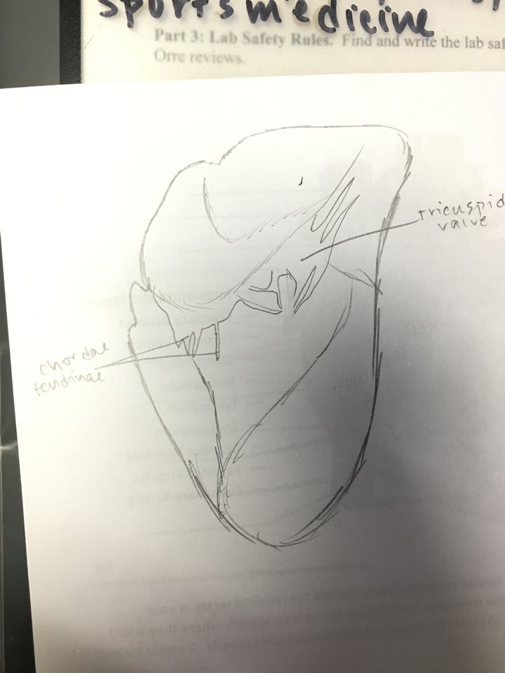
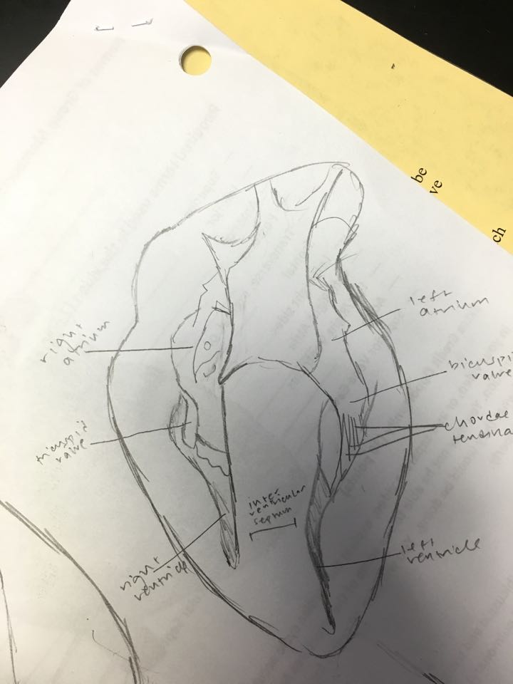
5. The coronary sinus is a blood vessel that empties blood into the right atrium after receiving blood from the coronary veins. The inferior vena cava is a large vein that takes deoxygenated blood to the heart from the lower half of the body and empties into the right ventricle. The tricuspid valve is pictured as a flap between the right atrium and right ventricle.
12243647_10206320091174248_233177747_n.jpg

7. The anchoring of the heart valves is so important because it ensures that the valves do not get carried away with the blood and are not a part of the backflow as well.
12231309_10206320091134247_1097553905_n.jpg
9. The semilunar valve prevents backflow to the heart.
10. A swelling in the feet and ankles if a valve disease occurs on the right side of the heart is because the blood is supposed to come up from the inferior vena cava which goes through the right atrium, the right side of the heart but with this disease, it means that there is a backflow because the valves are not working, causing swelling. If a valve disease occurred on the left side of the heart, there wouldn't be sufficient blood going to the muscles and body since the left side of the heart, containing the aorta, sends oxygenated blood to the rest of the body.
11. The chordae tendinae look fibrous and tendon-like and I can see them connecting to their respective valves. The valves look like flaps
11. The left and right sides of the heart have several differences. For example, the left side is bigger and has more myocardium because it pumps blood throughout the entire body, while the right side is not as muscular since it only pumps blood to the lungs
13.
12244201_10206320091054245_678089891_n.jpg
https://drive.google.com/file/d/0B20g3GH8iX_Gdm9nRE5PbkpGSGc/view?ts=564aab47

Wednesday, November 11, 2015

Unit 3 Reflection

This unit was all about the circulatory system, understanding the workings of the heart and some of its accessory organs to understand more about heart diseases and how to prevent and diagnose them. The cardiovascular system is quite complex, pumping heart all throughout the body, oxygenated and deoxygenated blood. The heart has four chambers and simply put, deoxygenated blood goes in to the right atrium and through the right ventricle to the lungs where it is 'oxygenated' then goes back through the heart through the left atrium then left ventricle and out to the rest of the body. To maintain a healthy cardiovascular system, exercise, a healthy diet, and much more are needed otherwise there can be plaque buildup in the arteries of the heart that can be life-threatening. Heart disease includes many cardiovascular diesases such as heart attacks, cardia arrenst, strokes and more which are caused by the atherosclerosis of coronary arteries. However to promote cardiovascular health, people can eat foods with less LDL cholesterol and more HDL cholesterol, exercise more, and not smoke. I would like to learn more about the treatments for cardiovascular diseases and how different things affect those treatments. 
Dissection of a sheep's heart
For this unit, I felt that it was easier because I could go through the heart and its chambers and veins to truly understand the process rather than just be memorizing what is good and what is bad. However through the heart dissection I also got to learn that real world application is important, especially in subjects such as anatomy because I found that the heart that we worked with was quite different from a heart in the anatomy textbook. In the last reflection I mentioned wanting to do research on sections that I did not understand as well or remember as well and I actually did that for some of the treatments and I think it helped me understand the concepts more thoroughly. For the rest of the semester, I would like to go over what I learned in class that day for homework as well rather than just waiting to study before the test. Like that chart that we were shown in class about how well people remember things, I think that I should apply that as well.

Tuesday, October 13, 2015

Measuring blood pressure

When measuring blood pressure, there are two numbers that must be taken: systolic pressure and diastolic pressure. The systolic pressure can be determined by where the meter is when you first hear a pulse beat while the diastolic pressure is determined by the number on the meter when the sound of the beat changes and blood flows better as the pressure is released more. To measure blood pressure, a sphygmomanometer and stethoscope is used while when measuring heart rate, either the fingers or stethoscope are used. However, using your thumb is not ideal when measuring heart rate since the thumb has a heart beat! Using a sphygmomanometer can actually be quite simple. First, wrap the cuff around a person's right bicep and find their heartbeat in or near the crook of their elbow. Before beginning, make sure the person has their palm facing up and arm propped up, around chest level. Then close the valve on the pump and pump the cuff until the pressure gauge reaches 150, then slowly release the air (by opening the valve). Watch the gauge and the number it is on when the first heartbeat is heard is the systolic pressure while the number on it when the sound of the heartbeat changes to a whooshing sound, is the diastolic pressure.

Displaying IMG_4936.JPG Displaying IMG_4935.JPG


Nicole
Cassi
Blood Pressure Test 1
205/60
100/58
Blood Pressure Test 2
94/60
104/62


Test 1 (Nicole)
Test 2 (Cassi)
Pulse rate (radial)
60
66
Pulse rate (carotid)
60
72
Stethoscope
66
72